Microplastics are present in aquatic environments, soil, sediments, food, consumer goods, and industrial processes. As research expands and regulatory expectations become more demanding, laboratories require analytical methods capable of accurately identifying polymers, counting particles, and determining particle-size distributions.
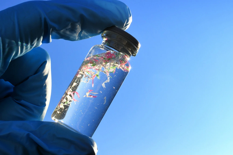
Image Credit: MargJohnsonVA/Shutterstock.com
Bruker provides microplastics researchers and testing laboratories worldwide with a broad portfolio of spectroscopic instruments, software, and workflows. Whether laboratories require rapid, high-throughput screening, submicron particle characterization, or reporting aligned with recognized standards, Bruker supports the selection of an analytical approach suited to the intended application.
Bruker’s solutions support reliable assessment of:
- Polymer composition
- Particle numbers
- Particle-size distributions
- Classification of shapes, fibers, and fragments
- Weathered, aged, and degraded polymers
- Samples containing multiple materials
- Regulatory reporting requirements
Microplastics can be detected and analyzed in:
- Drinking water, wastewater, and surface water
- Marine and ocean-related samples
- Soil and sediment
- Food and beverages
- Consumer products
- Industrial process streams
- Airborne particles
Frequently Asked Questions About Microplastics Analysis
This FAQ addresses key considerations in microplastics testing, including how to select a suitable method, what different regulations require, and the practical advantages and limitations of different approaches.
Microplastics: The Basics
What is microplastics analysis?
Microplastics analysis is the scientific examination of plastic particles smaller than 5 mm in environmental, biological, food, and water samples. The process may involve detecting, identifying, counting, and characterizing the particles. A complete assessment determines not only whether microplastics are present, but also their quantity, dimensions, morphology, and, most importantly, their polymer composition.
Where have microplastics been detected?
Microplastics have been reported in a wide variety of environments and biological materials, including deep-ocean sediments, seawater, Arctic ice, farmland soil, indoor air, bottled and tap water, human lung tissue, blood, and placental tissue. Their occurrence has been documented in marine and freshwater systems, atmospheric samples, terrestrial soils, food and drinking water, and biological specimens.
Choosing the Right Microplastics Analysis Method
What are the two primary approaches to microplastics analysis?
Microplastic analysis generally follows either a mass- or particle-based approach. Mass-based methods, including pyrolysis-GC/MS, determine polymer concentration by measuring mass. Because the sample is destroyed during analysis, these techniques cannot normally provide particle numbers, individual dimensions, or morphological information.
Particle-based techniques, such as IR and Raman microscopy, analyze individual particles directly. They can identify polymer composition while retaining particle-specific information, including particle count, size, and shape.
What are the three primary spectroscopic methods for microplastics analysis?
The three principal spectroscopic options are FTIR microscopy, IR laser imaging, and Raman microscopy. Each combines spectroscopic measurements with microscopy to evaluate the molecular vibration spectrum of a particle. However, the methods differ in areas such as sample compatibility, accessible spectral range, measurement speed, and spatial resolution.
How do FTIR, IR laser imaging, and Raman microscopy differ?
FTIR microscopy is a well-established technique for routine microplastic characterization. By integrating infrared spectroscopy and microscopy, it identifies polymer types while also supplying information about particle dimensions and morphology. The method is particularly suitable for automated filter examination and applications requiring consistent, routine particle analysis.
IR laser imaging microscopy (ILIM) also relies on infrared spectroscopy, but uses tunable quantum cascade lasers (QCLs) to perform rapid hyperspectral imaging. Its principal benefit is measurement speed. Large filter surfaces can be chemically mapped considerably faster than with conventional FTIR, which makes ILIM well-suited to high-throughput measurements and complete-filter analysis.
Raman microscopy uses visible or near-infrared excitation instead of mid-infrared illumination. The shorter wavelengths provide greater spatial resolution, making Raman especially useful for small particles, including those near 1 µm and, when measurement conditions are favorable, those below that size. Raman measurements may also reveal information about pigments, fillers, and inorganic constituents.
Particle Identification and Detection Limits
Why is chemical identification needed in microplastics analysis?
Appearance alone is generally insufficient to distinguish a synthetic polymer particle from naturally occurring materials such as cellulose or mineral fragments. Establishing this distinction can be important when investigating particle sources and origins. Chemical identification through spectroscopic methods confirms polymer identity and separates plastic particles from natural or mineral materials with similar visual characteristics.
What are detection limits and where do they come from?
Diffraction places a fundamental limit on the spatial resolution of optical microscopy, meaning that the illumination wavelength strongly influences the smallest features that can be resolved. Since mid-infrared radiation has micrometer-scale wavelengths, FTIR microscopy and QCL-based IR imaging both encounter an inherent resolution limit as particle dimensions decrease.
In routine microplastic applications, IR spectra that are suitable for interpretation can generally be obtained from particles in the low-micrometer range. At approximately 5 µm, however, dependable identification of individual particles becomes progressively more difficult because the particle size approaches the measurement’s spatial-resolution limit.
Raman microscopy is often more effective for smaller particles. Its use of much shorter visible or near-infrared wavelengths results in significantly higher spatial resolution. Consequently, particles around 1 µm and, under appropriate conditions, particles smaller than 1 µm may be characterized.
What are nanoplastics and how are they evaluated?
Nanoplastics are commonly defined as plastic particles measuring less than 1 µm. Analysis at this scale is especially challenging because particles may approach or fall below the resolving power of standard optical techniques.
Raman microscopy is therefore frequently considered for very small particles. Further characterization within the nanoscale may require more specialized approaches, including enhanced Raman methods and other analytical techniques developed for nanoscale measurements.
Throughput, Full-Filter Analysis, and Subsampling
What is subsampling in microplastics analysis?
Subsampling means measuring only a selected, supposedly representative part of a filter or sample instead of examining the complete filter. This practice is often used to reduce acquisition time in particle-based spectroscopic workflows, particularly when a sample contains a large number of particles.
However, particles may not be distributed evenly across a filter. As a result, subsampling can introduce uncertainty caused by spatial bias, losses during handling, particle crowding, or an insufficient number of measured particles.
Why can subsampling be risky in microplastics analysis?
Although subsampling reduces measurement time, the selected area may not accurately reflect the composition of the entire filter. Particle clustering, edge effects, uneven deposition, and handling-related losses can influence the reported particle count, polymer distribution, and particle-size profile.
Bruker’s full-filter IR imaging workflows eliminate this additional sampling step. ILIM can examine complete 25 mm filters in less than 30 minutes, including spectral collection and automated processing. This supports high-throughput analysis without the uncertainty associated with subsampling.
Sample Preparation and Filter Selection
Why is sample preparation so valuable in microplastics analysis?
Sample preparation is often the largest contributor to differences between laboratories. If organic and inorganic matrix components are not adequately removed, the resulting data may contain false identifications or otherwise unreliable results. Software can recognize and classify a certain amount of contamination, but this capability has limits.
When matrix material covers or conceals microplastic particles, software cannot recover information that was never successfully measured on the filter.
What filter should be used for microplastics analysis?
The appropriate filter depends on the selected measurement technique. Anodisc (aluminum oxide) filters are frequently used for IR transmission measurements. Silicon membrane filters offer broad access to the mid-IR region and are also compatible with Raman analysis.
Gold-coated polycarbonate filters are well-suited for Raman measurements and IR transreflectance. PTFE, metal mesh, and nitrocellulose filters can impose significant limitations in imaging workflows, although they may remain appropriate for single-point ATR measurements.
Can the same filter be used for both IR and Raman analysis?
Silicon membrane filters are compatible with both IR transmission and Raman measurements. Gold-coated polycarbonate filters can also be used for Raman and IR transreflectance analysis. However, anodisc filters are generally unsuitable for Raman because their surface becomes rough at high magnification and may produce fluorescence under laser illumination.
Data Quality and Classification
How is spectroscopic data evaluated in microplastics analysis?
For imaging-based methods, automated software such as Bruker's MPID converts millions of measured spectra into a particle list. This list can include the identity, dimensions, shape, and number of each detected particle, saving users the arduous task of manually reviewing such a large volume of data.
Automated classification can provide greater robustness than conventional library matching, particularly when the sample contains contamination or weathered and environmentally aged particles.
Why does classical library matching fail for environmental microplastics?
Environmental particles may be altered by contamination, chemical weathering, and ultraviolet exposure. These processes modify their spectral signatures, causing them to differ from clean reference materials in ways that basic template matching may not reliably accommodate. When conventional library matching is insufficient, a neural network trained using spectra from real-world degraded particles can help preserve classification precision.
What is a confidence score (HIT score) in microplastics classification?
A confidence score, also called a HIT score, expresses how closely the spectral feature vector of a measured particle corresponds to an authenticated reference spectrum for a particular polymer class. Analysts can define an acceptance threshold and retain only identifications that meet the required confidence level.
This threshold reflects a balance between sensitivity and specificity. A lower threshold may identify more potential particles but can also increase the number of incorrect assignments. A higher threshold is more selective and reduces false classifications, although some weaker or altered matches may be excluded.
Regulatory Requirements and Reporting
Which spectroscopic techniques are recognized for regulatory microplastics analysis?
For microplastic testing in drinking water, Commission Delegated Decision (EU) 2024/1441 recognizes vibrational spectroscopy methods, including μ-FTIR, μ-Raman, and equivalent techniques such as QCL-IR. This allows laboratories to select an approach according to requirements for particle size, throughput, and the level of analytical information needed.
Which regulations govern microplastics analysis in water?
The main regulatory and standardization references include EU Directive 2020/2184, which establishes the framework for drinking water in the European Union; Commission Delegated Decision (EU) 2024/1441, which specifies vibrational spectroscopy methods including μ-FTIR, μ-Raman, and equivalent approaches such as QCL-IR; ISO 24187:2023, which sets out general principles for microplastics analysis across environmental matrices; and ISO 16094-2:2025, which covers vibrational spectroscopy for drinking water and water containing low concentrations of suspended solids.
ASTM WK87463 is currently under development as a test method for identifying and quantifying microplastics in water using IR spectroscopy.
Do regulations require FTIR or Raman methods?
EU Commission Delegated Decision (EU) 2024/1441 does not prescribe one exclusive vibrational spectroscopy technique. It permits methods such as μ-FTIR, μ-Raman, and equivalent technologies, including QCL-IR (IR laser imaging), for the compositional analysis of microplastic particles and fibers.
The best choice depends on factors such as particle dimensions, sample composition, throughput requirements, and the intended analytical criteria.
What is the difference between ISO 24187 and ISO 16094?
ISO 24187:2023 establishes broad principles and minimum requirements for microplastics analysis in environmental matrices. ISO 16094-2:2025 provides more specific guidance for particle-based analysis using vibrational spectroscopy in drinking water and water with low suspended-solids content. Laboratories examining these types of water samples should therefore consider the requirements of the applicable standards.

This information has been sourced, reviewed, and adapted from materials provided by Bruker Optics.
For more information on this source, please visit Bruker Optics.