Electron microscopes manipulate the relatively short wavelength of electrons (100,000 times shorter than the photons that make up visible light) to study the structures of microscopic objects. They create magnetic fields shaped into an electron-optical lens, which works analogously to a conventional optical microscope’s glass lens. This electron-optical lens can magnify up to 10,000,000 times with a resolution better than 50 pm (picometers, one trillionth of a meter).
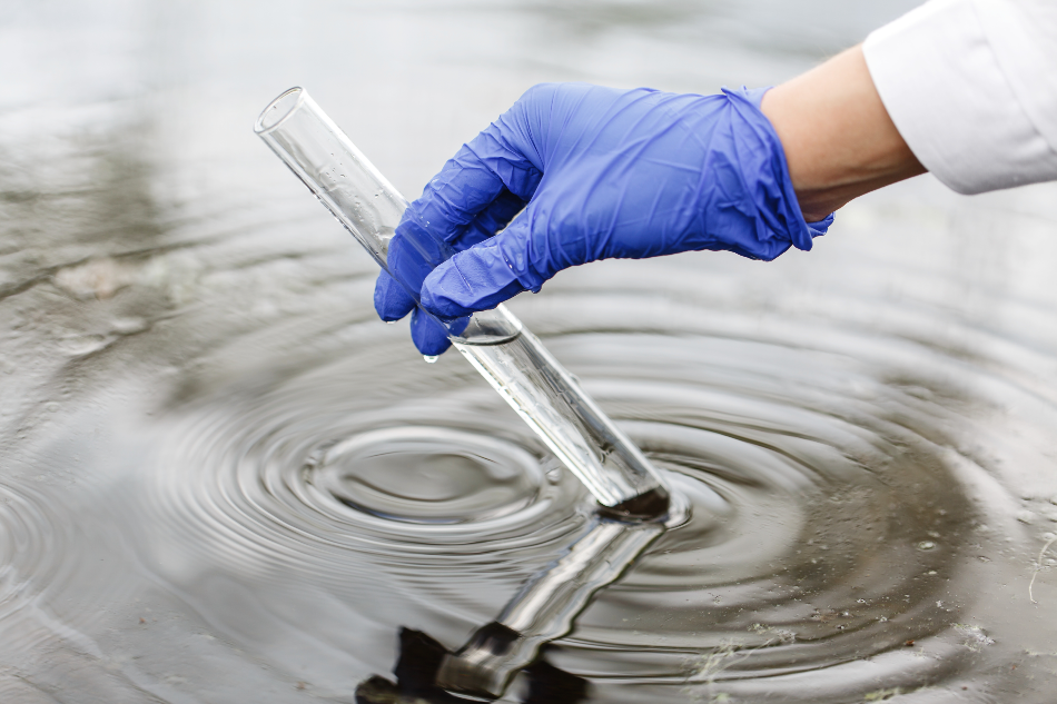
Image Credit: IVASHstudio / Shutterstock.com
Electron microscopy can be used to observe the ultrastructure of physical objects, which is the architecture of individual cells in the specimen. This has a range of applications in numerous fields, including semiconductors and data storage, biology, materials research, and various industries that rely on the precise understanding and detection of minuscule parts of component materials. Aquatic sciences also frequently use electron microscopy, especially in studying contaminants in water.
Understanding How Beaches Influence Long-Term Oil Pollution
The Prestige oil tanker sank off the coast of Galicia, Spain, in 2002, spilling 77,000 tonnes of oil into the ocean and thereby contaminating the local seawater and beaches. To understand how oil contamination affected the changing landscapes of beach sands, a team of researchers from the Universities of Vigo (Spain) and Montpelier (France) sampled seawater samples from different areas using electron microscopy to identify the levels of oil they contained (Fernández-Fernández et al., 2011).
Finding Invisible Viruses in Seawater
A University of Maryland team used transmission electron microscopy to reveal prophages, invisible viruses that become part of the microbacterial ecosystem by integrating their DNA into hosts in seawater (Chen et al., 2006). In transmission electron microscopy, the beam is projected through the specimen to generate the magnified image. The strategy demonstrated incorporates genome research with traditional microbiological techniques to find these invisible viruses with electron microscopy.
Testing Denitrification of Irrigation Water
Electron microscopy has also been used to test a new method for removing nitrates from synthetic infiltrate used in agriculture. In the method, methanol bacteria are added to the water, which significantly affects nitrate levels in soil irrigated by the water. Electron microscope images of soil samples supported the researchers’ findings that nitrates were removed from the water by adding methanol bacteria (Siripattanakul et al., 2010).
Identifying Potentially Toxic Microalgae and Poisoning Acid
Another University of Maryland team used electron microscopy to identify potentially toxic microalgae species in water in the Chesapeake Bay, an estuary in the northeast United States. The researchers used transmission electron microscopy to study samples of water. They found six species of Pseudo-nitzschia – potentially toxic diatom microalgae – and found information on domoic acid levels in the water. Domoic acid is a neurotoxin and the cause of amnesic shellfish poisoning, which can harm humans and other predatory animals when they eat shellfish, sardines, and anchovies in which it has accumulated.
Using This Research to Avoid Contaminations in Water
The examples above all use electron microscopy to better understand how waters around the world can be contaminated by human materials and pollutants or by microscopic bacteria. In each case, and in many others, the electron microscope’s ability to allow researchers to observe, identify, and study cell-sized organisms has enabled a clearer view of water contamination. This better understanding ultimately leads to new ways of avoiding contamination.
In some cases, such as the denitrified water, electron microscopy can be used to test novel decontamination techniques, such as adding bacteria to water to prevent it from contaminating the soil.
References and Further Reading
Chen, F., Wang, K., Stewart, J. and Belas, R. (2006). Induction of Multiple Prophages from a Marine Bacterium: a Genomic Approach. Applied and Environmental Microbiology, 72(7), pp.4995–5001.
Fernández-Fernández, S., A.M. Bernabeu, F. Bouchette, D. Rey, and F. Vilas. "Beach Morphodynamic Influence on Long-term Oil Pollution: The Prestige Oil Spill." Journal of Coastal Research, 2011, 890-93. https://www.jstor.org/stable/26482301
Siripattanakul, Sumana, Carlee J. Pochant, and Eakalak Khan. "Nitrate Removal from Agricultural Infiltrate by Bioaugmented Free and Alginate Entrapped Cells." Water Environment Research 82, no. 7 (2010): 617-21. http://www.jstor.org/stable/27870353.
Disclaimer: The views expressed here are those of the author expressed in their private capacity and do not necessarily represent the views of AZoM.com Limited T/A AZoNetwork the owner and operator of this website. This disclaimer forms part of the Terms and conditions of use of this website.